Mr. A, a 60-year-old male, presented to the emergency department (ED) with progressive stridor. His history was notable for radical tonsillectomy, neck dissection, radial forearm free flap, and tracheostomy for a locally and regionally advanced p16+ squamous cell carcinoma (SCCA) approximately two months earlier. Mr. A’s tracheostomy had been removed 1 month prior at Penn Otorhinolaryngology-Head and Neck Surgery, and he had just begun postoperative chemoradiation.
A Penn otolaryngology consult resident was called for urgent airway evaluation, and following a consult with the attending faculty, and a subsequent conference call with the junior and senior residents, a plan for definite hospital admission and likely nasopharyngolaryngoscopy (NPL) was initiated.
Mr. A reported having progressive dyspnea since his decannulation a month before presentation, and his medical history included sleep apnea and chronic renal insufficiency. Primary assessment revealed that he had trismus, a well healed free flap in the lateral oropharynx, a Mallampati grade of three and a nearly healed tracheostomy site with a small amount of granulation tissue in the residual tract.
Complicating matters: Mr. A was considered a “PUI” (patient under investigation/rule out COVID-19). Although he had stridor and positional dyspnea (when lying down), he had a regular respiratory rate when calm, did not require supplementary oxygen, and was otherwise stable.
In the setting of a potential COVID infection, team members were at risk of exposure even during a standard head and neck exam. Thus, any airway procedure including NPL would require full personal protective equipment (PPE). To accommodate the added risks and resources needed for airway evaluation and management in a PUI, the Penn Airway Rapid Response (ARR) team mobilized key personnel and equipment should the situation deteriorate, and discussed options available with all key personnel.
It was decided that the consult faculty should don full PPE (powered air-purifying respirator [PAPR], gown, and gloves) to do an airway assessment. Upon their entry to the room, however, Mr. A (who was masked) became extremely anxious, and his stridor loudened. When notified of the need for NPL evaluation and that he would not be receiving topical anesthesia to avoid the risk of virus aerosolization and worsening airway, Mr. A became more anxious, had increasingly loud stridor and tachypnea.
A disposable bronchoscope and tracheostomy set, among other vital equipment, were prepared at bedside. In the ED negative pressure room, the treating faculty donned full PPE and prepared for intubation or tracheostomy in case of decompensation.
Mr. A was preoxygenated should a rapid sequence induction be needed for oral intubation. Fiberoptic NPL revealed mild laryngeal edema and normal vocal cord mobility, suggesting a subglottic or tracheal source of obstruction. Mr. A tolerated the procedure well, remained stable, and was weaned back to room air.
It was determined that it would be best to avoid further intervention (including a CT chest and trachea evaluation) pending COVID testing, which as subsequently performed and provide negative.
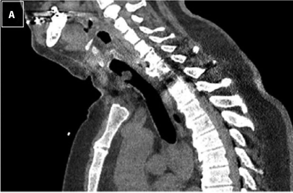 Figure 1A: Representative CT image demonstrating pedunculated tracheal lesion.
Mr. A was masked and transferred to a negative pressure room in the surgical ICU, where airway equipment was again placed at bedside. A chest CT obtained without contrast (a nod to Mr. A’s history of renal insufficiency) revealed multiple pulmonary metastases, a subcarinal mass (likely nodal), slight narrowing of the subglottis, and a mass in the trachea near the prior tracheostomy site, felt to be most consistent with a granuloma (Figure 1A).
Mr. A remained relatively stable but was in need of airway intervention. Given the negative COVID test, clinical picture with absence of fever, and the likely structural source of airway distress on imaging, it was decided Mr. A could be taken to the operating room without the need for a negative pressure room or PAPRs. Per current hospital guidelines for aerosolizing procedures (even in presumed COVID-negative patients), all staff wore N-95 masks, face shields, gowns, and gloves.
Following a thoracic anesthesiology consult, a rigid and flexible bronchoscopy was prepared, as well as possible emergent tracheostomy, and the team proceeded with a rapid sequence induction using videolaryngoscopy. The anesthesiologist had a grade I Cormack and Lehane view with the C-Mac videolaryngoscope and was able to pass an 8.0 endotracheal tube (ETT) through the glottis.
Once ventilation was confirmed, a flexible bronchoscope was passed through the ETT and visualized the tracheal lesion. The team was then able to carefully pass the tube over the bronchoscope beyond the soft mass. The table was turned 90° and the patient suspended for telescopic laryngoscopy with an excellent view using a Lindholm laryngoscope.
Ventilation was held, the ETT cuff was deflated, and the tube was withdrawn, revealing a pedunculated, friable mass which was then removed with upbiting cupped forceps and sent to pathology. Bleeding was controlled with topical oxymetazoline cottonoids. With the airway now clear and Mr. A now known to be an easy intubation via direct laryngoscopy, the decision was made to extubate.
Mr. A had immediate resolution of stridor and dyspnea and was discharged the following day. Pathology of the tracheal mass revealed carcinoma with similar appearance to his tonsil cancer, strongly suggesting metastatic disease involving the anterior tracheal wall.
This case appeared in a slightly different format in Head & Neck 2020;42:1273–1277.
Discussion
The COVID-19 pandemic has complicated the management of head and neck cancers, particularly for patients with underlying immunosuppressive disorders. The imposition of protective precautions has tasked head and neck oncologists with the need to temper the risks of cancer progression while managing the risks implicit in increased susceptibility to life-threatening complications from viral exposure.
Necessary head and neck cancer surgeries continued at the Department of Otorhinolaryngology-Head and Neck Surgery in the days and weeks after the onset of the COVID pandemic in Philadelphia. The treatment of these complex patients required not only depth of experience, but an evolving algorithm defined by on the spot innovation, rapid decision-making and truncated response times.
When originally published in a slightly different format, the three cases in this series were intended at publication to provide guidance to clinicians struggling with how to best counsel and manage this unique subset of patients under the difficult circumstances of the COVID era.
Read other complex cases for head and neck cancer treatment during COVID
Management of Complex Head and Neck Cancers in the COVID Era – Case #2
Management of Complex Head and Neck Cancers in the COVID Era – Case #3