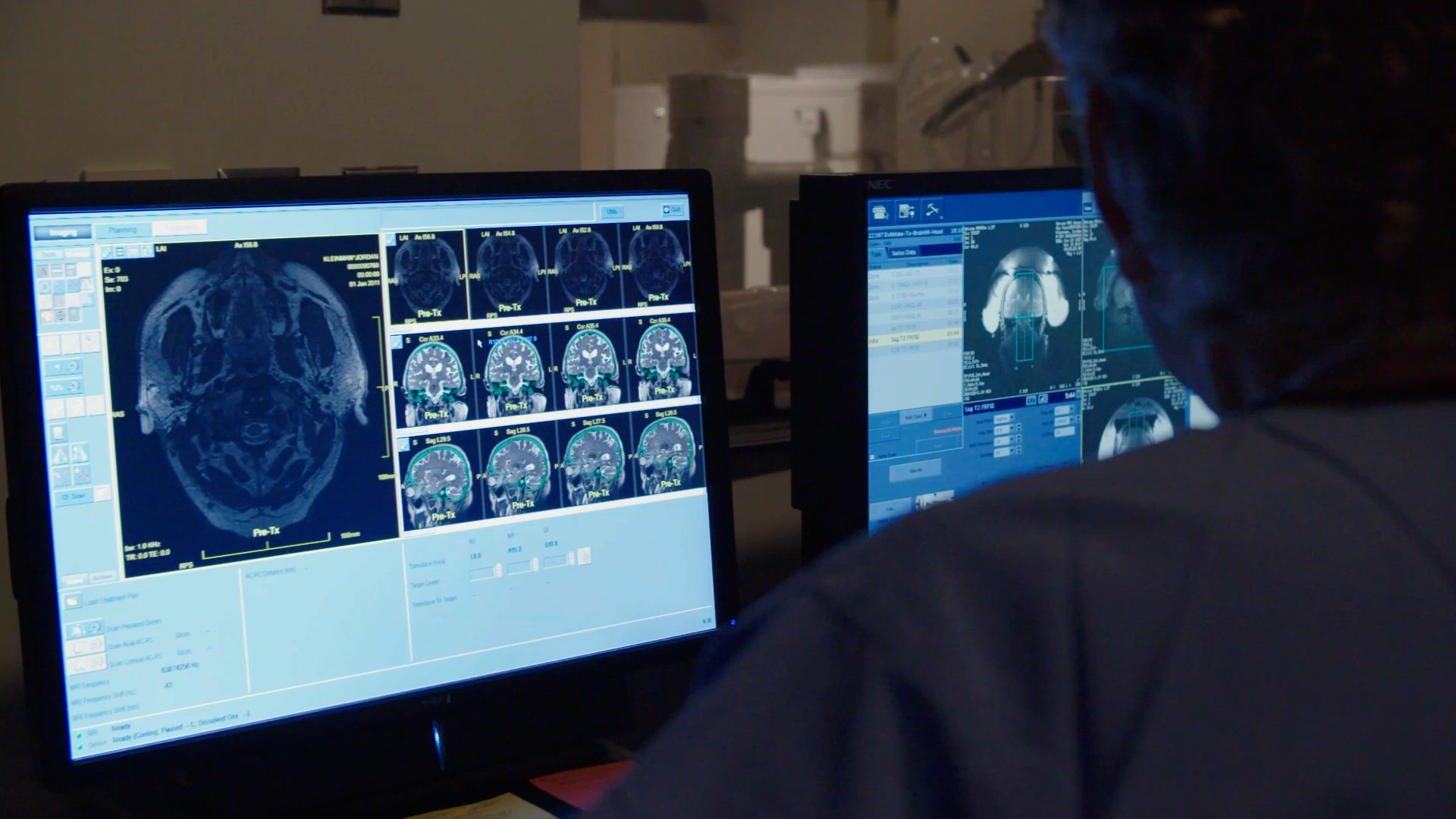
Intraoperative magnetic resonance imaging (iMRI) is a useful technique for guiding neurosurgeons when removing brain tumors and treating epilepsy. Using a combination of a magnetic field and radio waves, iMRI is used to get real-time images of the brain for monitoring brain activity; watching for any bleeding or clots that may arise; protecting brain functions; and preventing damage to surrounding tissues and nerves. For neurosurgeons treating tumors, it is also used to confirm the entirety of the tumor has been resected.
Although multiples images are taken prior to operating, using iMRI during brain surgery has multiple benefits. Such as checking to see if the brain has shifted at all during the procedure, confirming what is abnormal or normal brain tissue, and making sure critical areas of the brain are being protected.
Utilizing iMRI makes complicated surgeries easier and faster when you're able to look at imaging within the same area that you're performing the surgery. This is made possible with the newly designed hybrid operating rooms at the Pavillion. At other medical centers, you'll often see surgeons having to move the patient back and forth between the operating room and a room with an available MRI.