
Images reconstructed from 20 projections for non-infarct sheep at 85 BPM Scan Time 48ms/slice (approx. 15 slices at 85 BPM), significant streak artifact low SNR.
 Images KWIC reconstructed from interleaved projections for non-infarct sheep at 85 BPM Scan Time 48ms/slice. Removal of streak artifact improved SNR, maintained temporal resolution.
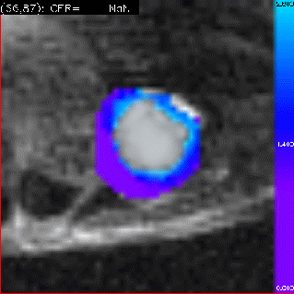
 Baseline contrast enhancement ratio (CER) generated for the left ventricle from first pass perfusion images. Demonstrates homogeneous profusion across the myocardium for all regions.
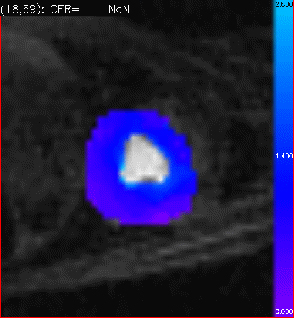
 Contrast enhancement ratio (CER) for the same sheep two weeks post infarct. Regions of hypoperfusion are seen in the left ventricular free wall where a transmural infarct is indicated. Non-transmural infarction is demonstrated in the border regions adjacent to the main infarct.