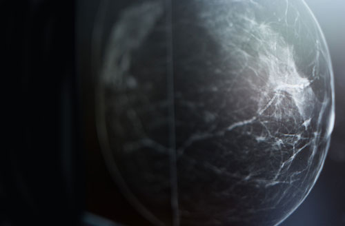
As a leader in breast clinical care and imaging research, the Breast Imaging Division of the Department of Radiology strives to enhance our patients' well-being through the delivery of the latest medical and scientific advances delivered with skill, integrity, respect and passion.
Collaborative, Research-Supported Clinical Care
The clinical mission of the Division of Breast Imaging is to provide individually tailored comprehensive evaluations for the early detection and diagnosis of breast diseases. As such, faculty members offer a highly personalized approach to breast imaging with an attention to patient education and comfort. The division's highly skilled staff uses technologically advanced diagnostic equipment that incorporates the latest in research developments to provide state-of-the-art breast imaging services and works in close collaboration with other departments to expedite patient care.
Accredited by the American College of Radiology, we maintain close clinical and research collaborations with the departments of Surgery, Radiation Oncology and Medical Oncology.
Equipment includes:
- Digital breast tomosynthesis units
- Dedicated breast ultrasound machines
- Prone digital stereotactic core biopsy units
- Dedicated breast MRI unit
Penn Radiology provides our patients breast imaging services, including digital breast tomosynthesis and breast MRI. The division is fully equipped for all forms of interventional procedures including stereotactic and tomosynthesis-guided, ultrasound, and MR guided biopsies. Since the center is a large referral center, the number of diagnostic studies and the volume of biopsies is quite high. In addition, there are very close clinical and research collaborations with the departments of Surgery, Radiation Oncology and Medical Oncology.
Versatile Educational Opportunities
Residents rotating through the breast imaging services are exposed to the highest quality of breast imaging with state-of-the art technology. Because of the large volume and complex cases, there is ample opportunity to learn all aspects of screening and diagnostic breast imaging. There is also the opportunity for in-depth exposure to a wide variety of interventional breast procedures performed under mammographic, ultrasonographic, or MR guidance.
The Breast Imaging Fellowship is a year of multi-modality breast imaging including tomosynthesis, breast MR and ultrasound. Because the section is a large regional referral site, there is a varied and complex daily clinical case load. Fellows are involved in daily, “at-the-view-box” teaching of the residents and are participants in the resident lecture series. There are also opportunities to rotate through the breast pathology, surgery, and medical oncology services.
The active basic science and clinical research programs offer unlimited opportunities to be involved in the many ongoing prospective research projects or in smaller retrospective data analyses. Graduates of the division’s fellowship have all been extremely successful in securing jobs in either academic or private practice after completion of this very well-rounded year.
Working to Improve Outcomes through Research
The section’s ongoing research includes multimodality breast imaging (digital breast tomosynthesis, ultrasound, MR, and PET imaging) for the screening, diagnosis, and cancer staging. Among the areas currently under study are:
- Correlation and quantification of breast density and complexity (as measured by mammography and MR) with clinical outcomes and breast cancer risk
- Chemoprevention and the potential impact on breast density change and breast cancer risk
- MR in the evaluation of response to neoadjuvant chemotherapy
- Digital mammography and digital breast tomosynthesis
- Contrast-enhanced digital breast tomosynthesis